Online ISSN: 2189-0544 Print ISSN: 0037-1017
公益社団法人日本生化学会 The Japanese Biochemical Society
Journal of Japanese Biochemical Society 97(5): 626-645 (2025)
doi:10.14952/SEIKAGAKU.2025.970626

総説Review

多種多様な両親媒性αヘリックスMultifaceted roles of amphipathic α-helices in membrane systems

大阪大学蛋白質研究所膜システム生物学研究室Laboratory for Membrane Systems Biology, Institute for Protein Research, The University of Osaka ◇ 〒565–0871 大阪府吹田市山田丘3–2 ◇ 3–2 Yamadaoka, Suita-shi, Osaka 565–0871, Japan

発行日:2025年10月25日Published: October 25, 2025
HTMLPDFEPUB3

両親媒性αヘリックスは脂質結合モジュールの一つであり,真核細胞からウイルスに至るさまざまな生物種由来のタンパク質に存在する.短いものは10アミノ酸程度から長いものは100アミノ酸を超えるものまで存在するが,すべてに共通しているのは膜と結合した際のαヘリックスの形成と,その構造をとった際に現れる両親媒性という2点である.そのシンプルな特徴のため,アミノ酸配列情報のみでは機能的な両親媒性αヘリックスなのかどうか判断が非常に難しい.また,両親媒性αヘリックスは個別に解析されているものが多く,類似性や共通性など,一般化した議論がなされる機会はそれほど多くない.そこで本稿では,基本的な性質からその解析手法,生理機能などを可能な限り幅広い視点でまとめることを目指した.後半では著者が専門とするオートファジー関連分子で報告されている両親媒性αヘリックスを最新の知見を交えながら概説する.

1. 両親媒性αヘリックスとは?

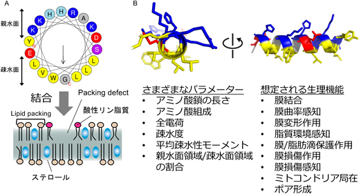
αヘリックスはタンパク質の二次構造の一つであり,アミノ酸主鎖のカルボニル基とアミド基の水素原子との間の分子内水素結合により形成される.らせん構造でアミノ酸残基の側鎖がヘリックスの外側を向いており,立体障害が最小になるように配置されている.1960年代に,ミオグロビンやヘモグロビン,アポリポタンパク質などに存在する多くのαヘリックスでは疎水性アミノ酸や親水性アミノ酸など,性質の似通ったアミノ酸残基が偏った分布を示すことが注目され始め,両親媒性αヘリックスの存在が認識されるようになった1).ここでいう両親媒性αヘリックスとは,親水性と疎水性のアミノ酸残基がαヘリックスに相対する配向をしており,全体として両親媒性を示すαヘリックスのことを指す(図1A).その後,Terwiligerらが,この両親媒性を評価するパラメーターとして平均疎水性モーメント(hydrophobic moment)を新しく定義したことにより,定量的な議論が可能になった2).このhydrophobic moment以外にも,アミノ酸鎖の長さ(chain length),全電荷(net charge),疎水度(hydrophobicity),親水面領域/疎水面領域の割合などのさまざまなパラメーターの違いから,これまでに多種多様な両親媒性αヘリックスが存在することが明らかになっている3).たとえば,アポリポタンパク質にはさまざまなタイプの両親媒性αヘリックスが存在しており,親水性側に存在する電荷を持つアミノ酸残基の数や分布のパターンに基づき,いくつかの異なるグループに分類されている3–5).その一方で,特定のアミノ酸残基の存在により両親媒性αヘリックスの機能が大きく変化することも知られている.グリシン(Gly)やプロリン(Pro)はαヘリックス構造を不安定にする性質からヘリックスブレーカーとも呼ばれるが,これらのアミノ酸残基を有する両親媒性αヘリックスも存在する.たとえば,曲率依存的に膜と結合する両親媒性αヘリックスにはGlyが比較的多く含まれており,構造的な柔軟性に寄与しているものと考えられる6, 7).また,膜損傷を誘導する一部の抗菌ペプチドにはProが存在し,Pro残基による折れ曲がり(kink)が膜損傷能を増強させる役割を果たしている8–10).両親媒性αヘリックスが結合する膜の膜脂質環境にも特徴があり,ホスファチジルエタノールアミン(PE)やホスファチジン酸(PA),ジアシルグリセロール(DAG)などの極性頭部が小さい脂質分子種が豊富な膜や,脂肪酸鎖の不飽和度が高い脂質分子によって形成された膜,曲率の高い膜構造などと結合する傾向がある.これは,両親媒性αヘリックスが結合する際に疎水性側のアミノ酸残基が脂質分子間に入り込むのに必要な空間(packing defect,図1Aレジェンドに解説)が多く,同じ両親媒性であるリン脂質分子から構成される生体膜と結合しやすい環境が生じることに起因する11–14)

Journal of Japanese Biochemical Society 97(5): 626-645 (2025)

図1 両親媒性αヘリックスは膜と結合することで機能する

(A)  両親媒性αヘリックスは,その構造により親水面と疎水面の両方を有し,全体として両親媒性の性質を示す.この特徴は生体膜を構成するリン脂質にも共通しているため,両親媒性αヘリックスは生体膜内の脂質分子間のすき間,すなわちpacking defect領域に入り込み,生体膜と結合する.packing defectという語は,脂質分子どうしが密接に並ぶ(lipid packing)構造に生じる欠陥(defect)に由来する.中央の矢印の長さは,両親媒性を表す疎水性モーメントの大きさを表示している.(B)両親媒性αヘリックスは,種類ごとにアミノ酸配列が異なっている.そのため,物理化学的なパラメーターもさまざまであり,生理機能も多岐にわたる.黄色:疎水性アミノ酸,青色:塩基性アミノ酸(リシン,アルギニン),水色:ヒスチジン,赤色:酸性アミノ酸(アスパラギン酸,グルタミン酸),灰色:側鎖が小さな疎水性アミノ酸(グリシン,アラニン),紫色:セリン,トレオニン.

2. 両親媒性αヘリックスを含むタンパク質の代表的なドメイン構造

では,どのようなタンパク質に両親媒性αヘリックスが存在するのか? これまでの研究報告を調べると,短いペプチドから膜タンパク質まで,さまざまなタンパク質に両親媒性αヘリックスが存在する(図2).以下に,タンパク質のドメイン構造パターンに従い,グループごとに分けて概説する(表1).

Journal of Japanese Biochemical Society 97(5): 626-645 (2025)

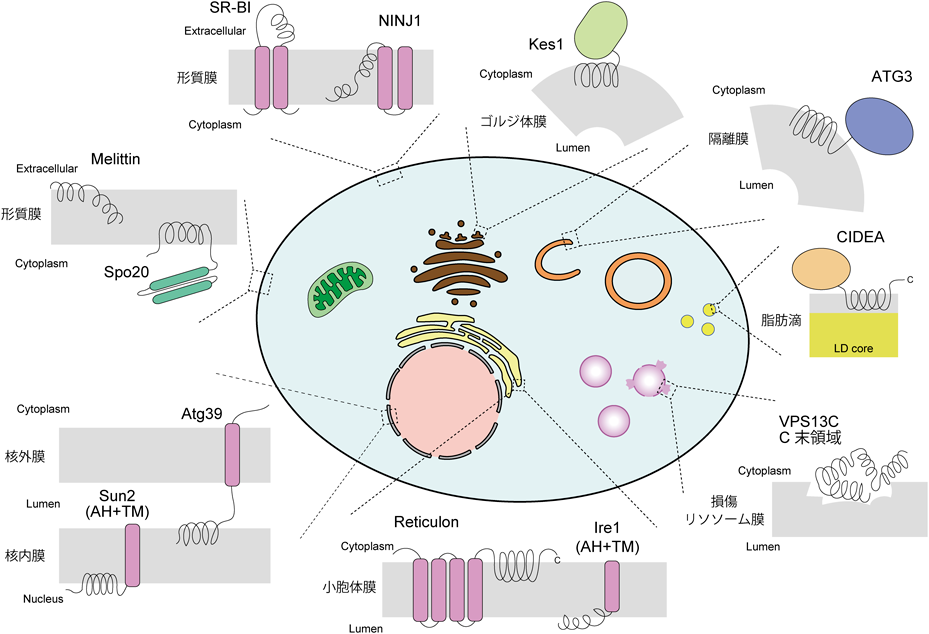
図2 さまざまな分子が両親媒性αヘリックスを有する

細胞表面や細胞内オルガネラに存在する両親媒性αヘリックスの例.両親媒性αヘリックスのみからなるものや,膜貫通領域を有するものなど,さまざまなものが存在する.(左上)形質膜上で,スカベンジャー受容体であるSR-BIの両親媒性αヘリックスは細胞外領域に存在する.パイロトーシスに必要な分子NINJ1は刺激に依存してオリゴマーを形成し,形質膜に穴を開けるが,その両親媒性αヘリックスは形質膜の外層と結合する.同様に,膜損傷を誘導するMelittinも形質膜の外層と結合する.一方で,Spo20の両親媒性αヘリックスは正に強く帯電しており,負に帯電したリン脂質が豊富な形質膜の細胞質側に結合する.(左下)核膜に局在する膜タンパク質のうち,両親媒性αヘリックスを有するものも存在する.オートファジー受容体の一つであるAtg39は核外膜に局在する膜タンパク質であり,その両親媒性αヘリックスは核内膜と結合する.また,核膜タンパク質であるSun2は核内膜に局在し,その両親媒性αヘリックスは核質側と結合する.(下)小胞体に局在するReticulonの両親媒性αヘリックスは細胞質側と結合するのに対し,Ire1の両親媒性αヘリックスは内腔側と結合する.(上)Kes1の両親媒性αヘリックスは,ALPSモチーフの一つであり,膜曲率の高い領域を認識して膜結合する.(右)CIDEAの両親媒性αヘリックスは,脂肪滴表面のリン脂質単層部分と結合する.(右下)四つの両親媒性αヘリックスからなるVPS13CのC末領域は,損傷したリソソーム膜に局在する.AH:両親媒性αヘリックス,TM:膜貫通領域.

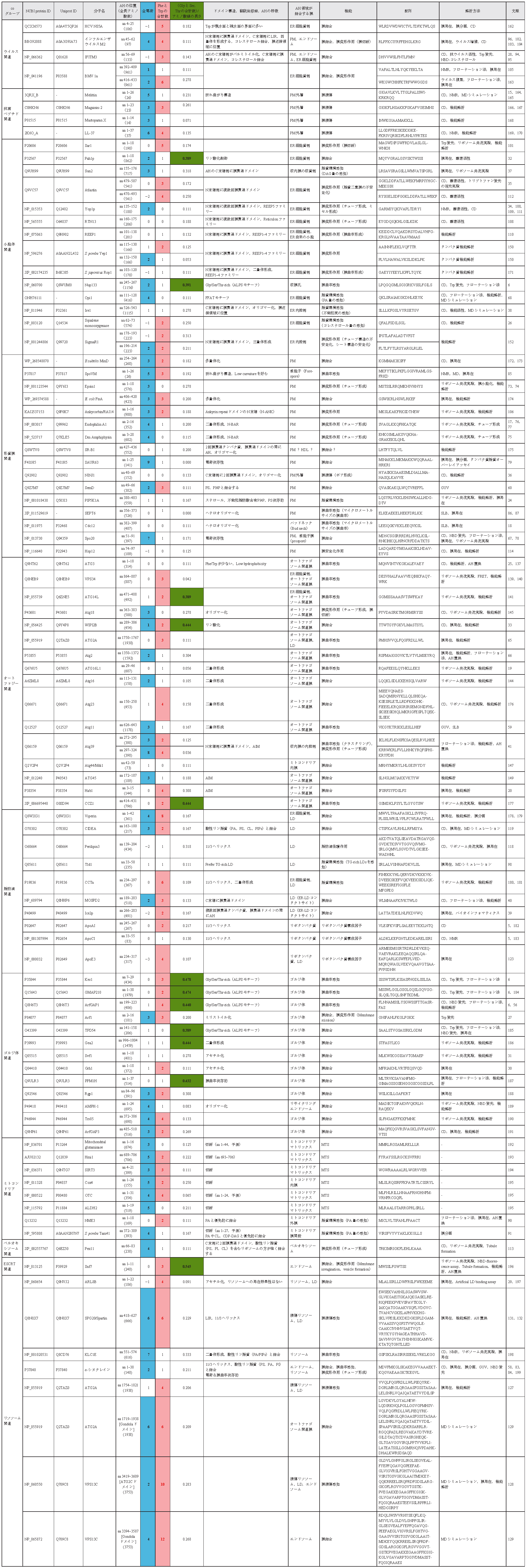
表1 代表的な両親媒性αヘリックス
これまでに報告のある両親媒性αヘリックスのうち,代表的なものを局在するオルガネラごとにまとめたもの.全電荷,PheとTrpの合計数,(GlyとSer, Thrの合計数)/アミノ酸鎖の長さなどのパラメーター値は,両親媒性αヘリックスの配列情報をもとにPythonを使って算出した25).グループによっては,いくつかのパラメーター値に特徴があることがわかる.たとえば,抗菌ペプチド関連やミトコンドリアターゲット配列関連,形質膜関連は,全電荷が正に帯電しているものが多い.ALPSモチーフは(GlyとSer, Thrの合計数)/アミノ酸鎖の長さの値が大きく,過去の報告どおり,これらのアミノ酸残基が多く含まれることを反映している6).リソソームの膜損傷を感知する両親媒性αヘリックスは,ほかのものと比べて配列が非常に長く,連続した両親媒性αヘリックスから構成される.ヒトATG3の両親媒性αヘリックスはオートファジー関連の中でも,全電荷が0であり,PheとTrpの合計数が少ないというユニークな特徴があることがわかる25).AH:両親媒性αヘリックス,PM:形質膜,ER:小胞体,LD:脂肪滴,MTS:ミトコンドリアターゲット配列,11/3ヘリックス:11アミノ酸あたり3回転するタイプの両親媒性αヘリックス,AIM:Atg8-interacting motif, LIR:LC3-interacting region, PS:ホスファチジルセリン,PA:ホスファチジン酸,PI:ホスファチジルイノシトール,PIPs:ホスホイノシチド,CL:カルジオリピン,DAG:ジアシルグリセロール,CDP-DAG:CDP-ジアシルグリセロール,TG-rich LDs:トリグリセリドが豊富に存在する脂肪滴,CD:円二色性スペクトル,NMR:核磁気共鳴,GUV:giant unilamellar vesicle, SLB:supported lipid bilayer, AH置換:AH部分を他のAHに置換したキメラ作製による機能解析実験.

1)両親媒性αヘリックスのみ

ハチ毒主成分のMelittinや抗菌ペプチドLL-37などの膜障害性ペプチドは,両親媒性αヘリックスのみからなる単純な構造である(図2左).大量のペプチドが膜と結合して深部まで入り込むことで,小さな穴を形成する.その結果,膜透過性が上昇し,致死的な膜損傷を引き起こす10, 15).これらの膜障害性ペプチドの多くは正に帯電していることから(表1,「抗菌ペプチド関連」を参照),細胞表面が負に帯電している細菌の細胞膜に優先的に作用することが知られている(Shai-Matsuzaki-Huangモデル)16)

2)可溶性タンパク質

膜損傷型の両親媒性αヘリックスを除くと,可溶性タンパク質の両親媒性αヘリックスは単独ではなく,ほとんどがほかのドメインやモジュールと連動して機能する.実際,可溶性タンパク質の多くは両親媒性αヘリックスを介して膜と結合すると同時に,他の分子と複合体を形成したり,またヘテロ/ホモ二量体や多量体を形成したりする(表1,「ドメイン構造」の列を参照)17–19).Atg8-interacting motif(AIM)やLC3-interacting region(LIR),FFATモチーフといった膜上に存在する分子と結合するドメインやモチーフを持つものも存在する20–22).これらの分子では,両親媒性αヘリックスを介した膜結合が,複合体の形成や多量体化により増強されたり,タンパク質間の相互作用と協調的に機能したりしていることが多い.

また,脂質代謝や脂質化反応に関わる酵素などでは,両親媒性αヘリックスが基質認識部位の近傍に位置している.膜上で生じる基質認識と酵素反応が密接に連動することで,基質依存的な酵素活性の制御や生成物/最終産物によるネガティブフィードバックなどが可能になっている23–26).両親媒性αヘリックス領域が翻訳後修飾を受けるものもあり,ミリストイル化やパルミトイル化,アセチル化などにより膜結合能が促進される(表1,「翻訳後修飾」の列を参照)27–31).一部の両親媒性αヘリックスでは,リン酸化により膜結合能が負に制御されることも明らかになっている32, 33)

3)膜貫通タンパク質

膜貫通タンパク質にも両親媒性αヘリックスを有するものが多く存在する(表1,「ドメイン構造」の列を参照).ヘアピン型膜貫通タンパク質Reticulonや2回膜貫通タンパク質Atlastinは小胞体膜に局在する分子であるが,いずれもC末側に両親媒性αヘリックスを有しており,小胞体膜の細胞質側(cytosolic face)と結合している(図2下)34, 35).形質膜に局在する2回膜貫通タンパク質であるNINJ1の場合はN末側に両親媒性αヘリックスを有しており,細胞膜の外層(extracellular face)と結合している(図2上)36).1回膜貫通タンパク質でも両親媒性αヘリックスを有しているものが存在する.たとえば,核膜タンパク質Sun2は核内膜(inner nuclear envelope)に局在し,膜貫通領域のN末側にある両親媒性αヘリックスが核質側(nuclear face)と結合する(図2左下)37).酵母Ire1の場合は,両親媒性αヘリックスが膜貫通領域と一部重なるユニークなタイプであるが,その領域を介して小胞体膜の内腔側(luminal face)と結合している(図2下)38).このように膜タンパク質では,自身が局在する膜と結合する場合が多い.ただし,結合する膜は必ずしも細胞質側に限定されず,内腔側や細胞膜外層など,さまざまな面から膜と結合する.

一方で,自身が局在する膜とは異なる膜構造と結合する両親媒性αヘリックスを有している膜タンパク質も存在する.小胞体膜に局在するIce2やMOSPD2は,両親媒性αヘリックスを介して脂肪滴と結合する39, 40).Atg39は核外膜(outer nuclear membrane)に局在する1回膜貫通タンパク質であり,その両親媒性αヘリックスは核内膜(inner nuclear membrane)の内腔側(luminal face)と結合する(図2左下)41).これらの分子は両親媒性αヘリックス依存的な膜結合を介して,オルガネラ膜が近接したコンタクトサイト領域で機能しているものと考えられる.

4)ミトコンドリア移行シグナル

ミトコンドリアマトリクスに輸送されるシグナル配列MTS(mitochondrial targeting signal)も両親媒性αヘリックスの性質を有しており,親水性側領域には正に帯電したアミノ酸残基が多い(表1,「ミトコンドリア関連」を参照)42–44).MTSはミトコンドリア外膜の膜透過装置であるTOM(translocator of the outer membrane)複合体の受容体であるTom20と相互作用し,ミトコンドリアへと標的化される.MTSをもつ前駆体タンパク質はその後,マトリクスへと到達し,MPP(mitochondrial processing peptidase)によってMTS部分が切断され,最終的に成熟タンパク質となる.MTSの正電化アミノ酸はTom20の近傍に位置するTOM複合体のサブユニットTom22によって認識されると考えられている.このように,これらの両親媒性αヘリックスは膜ではなく,Tom20/Tom22複合体と結合するシグナル配列として機能する45, 46).一方で,MTSがミトコンドリア外膜に直接結合することで前駆体タンパク質のアンフォールディングを促進するという報告もある47)

5)球状タンパク質とイオンチャネル

ミオグロビンやヘモグロビンなどの球状タンパク質は主にαヘリックスから構成される分子であるが,そのほとんどが両親媒性であることが知られている48).親水面を外側に,疎水面を内側に向けることで,エネルギー的に安定な構造をとっているものと考えられる.またアセチルコリン受容体などのイオンチャネルも両親媒性αヘリックスを有しており,疎水面を介して膜を構成する脂質分子と疎水性相互作用をしつつ,親水面を中心に向けることで,水溶性のイオン分子が通過するポアを形成する49, 50).最近では,両親媒性αヘリックスの領域を人工的に改変したイオンチャネルやナノポア技術への応用に関する報告もあり,注目されている51–53)

3. 両親媒性αヘリックスの解析手法

両親媒性αヘリックスを解析する代表的な手法を概説する.まず最も簡便な手法として,Gautierらによって開発されたHELIQUEST webserver54)に対象のアミノ酸配列をクエリとして入力し,両親媒性αヘリックスの候補領域を探索する方法がある.もし解析したいクエリ配列が多数存在する場合は,公開されているPython codeを利用すると解析を短縮できる25, 55).候補領域を絞ったら,次に実験に基づく検証を行う.一般的には,両親媒性αヘリックスの候補領域のペプチドを化学合成し,in vitroで実際に膜と結合するかどうか解析する.多くの両親媒性αヘリックスは水溶液中では構造をとらず,膜結合依存的にαヘリックス構造をとるため,リポソーム共存によるαヘリックス構造の増加を円二色性(circular dichroism:CD)スペクトルや核磁気共鳴(nuclear magnetic resonance:NMR)により解析する.また,αヘリックスの構造情報は得られないが,疎水環境依存的なトリプトファン蛍光特性の変化や蛍光色素NBDの蛍光強度の増加を利用したメソッドは定量性が高く,膜結合能の評価方法としては非常に有用な手法である6, 56).異なる炭素位にドキシル基を持つPCを用いたトリプトファン蛍光の消光実験により,両親媒性αヘリックスが膜結合時に,どの程度の深さまで挿入されているかを評価することもできる35, 57).合成ペプチドが凝集してしまい,可溶化できないこともあるので,そのような場合はタンパク質ドメインの全長を用いた解析が行われる.両親媒性αヘリックス領域を含むリコンビナントタンパク質を精製し,リポソーム共存下でのco-sedimentation(リポソーム共沈実験)やmembrane flotation(フローテーション法)などにより,膜結合画分を生化学的に解析することで評価する6, 56).giant unilamellar vesicle(GUV)やsupported lipid bilayer(SLB)を用いた手法もあり,GFPなどの蛍光タンパク質を付加したリコンビナントタンパク質とインキュベートし,光学顕微鏡により観察する.これらの手法では膜結合能だけでなく,膜変形作用なども評価することができる58–60).また,トリオレインで作製した人工脂肪滴と両親媒性αヘリックスとの結合を解析する手法も開発されている61–64)

一方で,これらは人工膜や人工脂肪滴を使ったin vitroでの解析であり,生体内に存在する複雑な膜脂質環境を完全には反映していない可能性がある.そこで,細胞内での膜局在を指標にするというアプローチも頻繁に用いられる.具体的には,両親媒性αヘリックスを含むタンパク質(野生型)と,その両親媒性αヘリックスの疎水性側領域にアラニン(Ala)や親水性アミノ酸を導入したもの(変異体)をGFP融合タンパク質として発現させて,両親媒性αヘリックス依存的な膜局在への影響を調べる65, 66).解析対象によっては,両親媒性αヘリックス領域のみで解析する場合,その膜結合能が弱く,十分な膜局在能が観察されないこともある.そのような場合はコイルドコイル領域を付加し,人工的に二量体化させるという方法も有用である24, 67)

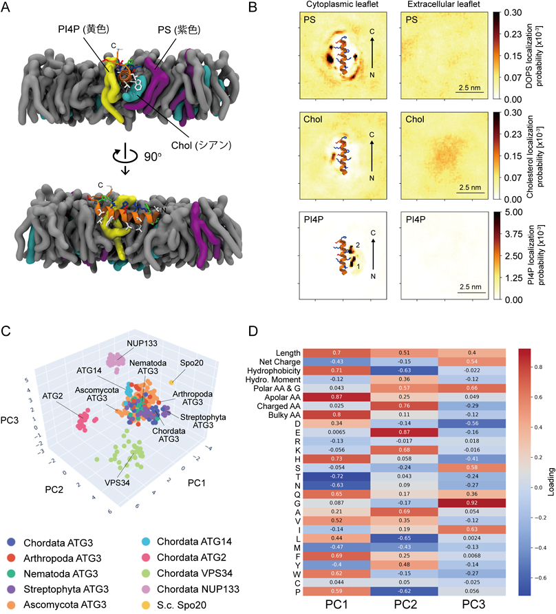
分子動力学(molecular dynamics:MD)シミュレーションによる両親媒性αヘリックスの解析も盛んに行われており,膜上でのダイナミックな挙動や,脂質分子と結合するアミノ酸残基の予測など,詳細な情報が得られるようになってきている14, 24, 38, 68, 69).計算に基づくin silico解析ではあるが,特定の脂質分子とアミノ酸残基との距離に関する情報やその時間依存的な変化を解析できるという点は,他の手法にはない強みである(図3A, B).

Journal of Japanese Biochemical Society 97(5): 626-645 (2025)

図3 両親媒性αヘリックス解析法の具体例

(A)ホスファチジルイノシトールリン脂質PI4,5P2合成酵素PIP5Kの両親媒性αヘリックス領域と脂質膜が結合する様子を全原子MDシミュレーションで解析したもの.黄色はホスファチジルイノシトールリン脂質PI4P, 紫色はPS, シアンはコレステロール(Chol),灰色はホスファチジルコリン,中央のオレンジはPIP5Kの両親媒性αヘリックスを示す.PIP5Kの両親媒性αヘリックスがPI4PとPSに挟まれる形で結合し,その際に生じる小さな隙間をコレステロールが埋める.PIP5Kの両親媒性αヘリックスのトリプトファン残基がコレステロールと近接した場所に位置していることが確認できる.(B)全原子MDシミュレーション解析における,PIP5K両親媒性αヘリックス周囲での各脂質の出現頻度.PI4PやPS, CholがPIP5Kの両親媒性αヘリックスの周囲に限局して分布している様子がわかる.(A, B)で用いている図は,文献24より一部改変して転載.(C)主成分分析による両親媒性αヘリックスの解析例.さまざまな生物種のオートファジー関連分子が有する両親媒性αヘリックスの物理化学的パラメーターをPythonにより算出し,主成分分析を使って次元削減.ATG3由来の両親媒性αヘリックスが中央に集中して分布していることが確認できる.Nup133はALPSモチーフの一つであり,Spo20は正に強く帯電した両親媒性αヘリックス.これらの両親媒性αヘリックスとは異なる分布を示していることから,ATG3由来の両親媒性αヘリックスがこれらとは異なるユニークな性質を有することがわかる.(D) Cにおける主成分分析のPC1, PC2, PC3の主成分負荷量(Loading)を示したもの.各物理化学的パラメーターやアミノ酸組成がどの程度寄与しているのか,その重みを表している.この表から,ATG3由来の両親媒性αヘリックスはかさの大きな疎水性アミノ酸残基が少なく,疎水性が低いという特徴が共通してみられることがわかる.Length:アミノ酸鎖の長さ,Net Charge:全電荷,Hydrophobicity:疎水度,Hydro. Moment:疎水性モーメント,Polar AA & G:極性アミノ酸残基とGlyの合計数,Apolar AA:疎水性アミノ酸残基の数,Charged AA:電荷を帯びたアミノ酸残基の数,Bulky AA:PheとTrpの合計数.(C, D)で用いている図は,文献25より一部改変して転載.

最近では,機械学習を用いた両親媒性αヘリックスの解析も進んでいる.両親媒性αヘリックスはアミノ酸鎖の長さが比較的短いものが多いため,ホモログ分子でも生物種が異なると配列類似性が著しく下がってしまい,共通した特徴を見いだしにくい.そのような場合には,主成分分析などの教師なし機械学習が効果的であり,特定の分子の両親媒性αヘリックスに共通して保存されている特徴量を見いだすことができる(図3C, D25).また,深層学習を用いた両親媒性αヘリックスの予測アルゴリズムも開発が進んでおり70, 71),最近報告されたMemBrain(http://www.csbio.sjtu.edu.cn/bioinf/MemBrain/)などを使うことで,以前よりも効率的に両親媒性αヘリックスの候補領域を絞ることも可能になってきている72)

4. 両親媒性αヘリックスの生理機能

両親媒性αヘリックスが果たす役割は多岐にわたるが,基本的な機能はいずれも膜結合に起因する(図1).しかし,その膜結合が生じる分子メカニズムの違いや結合強度の強弱によって,膜への影響はさまざまである.ここでは,これまでに報告されている生理機能をいくつかの代表的なグループに分けて概説する.

1)電荷依存的な膜結合

膜融合に関わるSNARE分子の一つであるSpo20やエンドサイトーシスに関与するEpsin1, N-BARドメイン(Amphiphysin, Endophilin A1),オーキシン初期応答因子SAUR63などの両親媒性αヘリックスは,いずれも形質膜と結合するが,疎水度が低く,正に強く帯電しているという特徴がある(表1,「形質膜関連」を参照).形質膜の細胞質側にはホスファチジルセリン(PS)やPA,ホスファチジルイノシトール4,5-ビスリン酸(PI4,5P2)など,負電荷を帯びた酸性リン脂質分子が多いため,電荷依存的な静電相互作用を介して膜と結合していると考えられる17, 67, 73–79)

2)膜曲率依存的な膜結合

膜曲率センサーとして機能する両親媒性αヘリックスは非常に多くのものが存在し,AntonnyとDrinらの先駆的研究により,その重要な役割が明らかにされてきた7, 80).COPI小胞のコートマータンパク質の膜からの解離には,ArfGAP1の膜曲率依存的なArf1-GTPに対するGTPase活性が関与しており81),ArfGAP1が有する両親媒性αヘリックスが膜曲率を感知し,GTPase活性に膜曲率依存性があることが明らかになった56).その後の解析から,GMAP210やKes1, Nup133など,他の分子にも膜曲率を感知する両親媒性αヘリックスが存在することが報告され,ALPS(ArfGAP1 lipid packing sensor)モチーフと命名された(図2上).これらのALPSモチーフはほかの両親媒性αヘリックスと比較して,親水面側に位置する塩基性アミノ酸の数が非常に少なく,その代わりにグリシン(Gly),セリン(Ser),トレオニン(Thr)の割合が多い(表1,「(GlyとSer, Thrの合計数)/アミノ酸鎖の長さ」の列を参照).その物理化学的な特徴から,静電相互作用ではなく,疎水面による疎水性相互作用に依存した弱い膜結合を示す.このような状況下においては,膜が曲がることによって形成されるpacking defectが結合に必要なエネルギー障壁を下げ,膜結合を促進しているものと考えられる6, 82).α-シヌクレインが有する両親媒性αヘリックスも同様に膜曲率依存的な膜結合を示すが,ALPSモチーフとは異なる性質を示し,膜との結合にはPSなどの負電荷を帯びた酸性リン脂質が必須である83).これはα-シヌクレインの両親媒性αヘリックスの親水面が弱く正に帯電しており,かつ疎水面領域のかさが小さいという物理化学的な特徴に起因するものと考えられる(表1,「α-シヌクレイン」を参照)84).一方で,曲率が低い膜構造と結合するものも存在し,Septin分子(SEPT6, Cdc12 etc)やSpoVMなどの両親媒性αヘリックスは直径がマイクロメートルサイズの膜と優先的に結合することが知られている18, 85–87).しかしながら,これらの両親媒性αヘリックスが曲率の低い膜に対して,どのように選択性を示しているのか,詳細な分子メカニズムはまだよくわかっていない.

3)脂質環境感知

脂質組成の変化を感知するセンサーとして機能する両親媒性αヘリックスもさまざまなものが存在する.酵母Opi1はFFATモチーフを介して小胞体膜タンパク質Scs2との結合により小胞体膜に局在しているが,両親媒性αヘリックスが小胞体膜の細胞質側に分布するPAの量を感知する脂質センサーとしての機能を果たしている68).Spo20の両親媒性αヘリックスは,選択的にPAと結合するという知見から78),PAに対する脂質プローブとして利用されることがある88, 89).最近では,ミトコンドリアの外膜に局在するNME3や内膜に局在するTam41由来の両親媒性αヘリックスがPAと優先的に結合することが報告されている90, 91).しかしながら,これらのPAと結合する両親媒性αヘリックスはin vitroではほかの酸性リン脂質とも結合する可能性があることから,選択的なPAプローブとしての利用には慎重な検討が必要と考えられる67)

生体膜上のpacking defectを感知する両親媒性αヘリックスも多く存在する.たとえば,核膜タンパク質Sun2の両親媒性αヘリックスはDAGの量依存的に生じるpacking defectを感知することで核膜と結合する.これによりSun2が核膜にとどまることが可能となり,プロテアーゼ依存的な分解を回避している37).また,小胞体ストレス応答に関与する酵母のIre1では,膜貫通領域の直前に位置する両親媒性αヘリックスが脂肪酸鎖の不飽和度の変化を感知することで,飽和脂質依存的な小胞体ストレス応答を誘導する38).前述のALPSモチーフも曲率依存的な膜結合だけでなく,packing defect依存的な膜結合を示すことも報告されている12, 14, 92)

コレステロールは主要な生体膜構成脂質成分の一つであるが,コレステロールが増加するとリン脂質分子間の隙間が埋まり,lipid packingが促進することが知られている93).このlipid packingの変化を利用して,小胞体膜上のコレステロール量の増加を感知する両親媒性αヘリックスも存在する26).その一方で,一部の両親媒性αヘリックスでは,ステロール依存的な膜結合能の増強が報告されている.たとえば,抗ウイルス作用を有するIFITM394, 95)やインフルエンザウイルスのM2タンパク質96, 97)が有する両親媒性αヘリックスはステロールとの直接的な結合が示されている.ホスファチジルイノシトールリン脂質キナーゼPIP5Kの両親媒性αヘリックスに関しては,PSやホスファチジルイノシトール4-リン酸(PI4P)などのほかの酸性リン脂質とともに,形質膜上のステロール量を感知している(図3A, B24).これらの両親媒性αヘリックスでは,フェニルアラニン(Phe)やトリプトファン(Trp)などのアミノ酸残基とステロールの結合が示唆されており,これらが膜上での両親媒性αヘリックスを安定化しているものと考えられる.

興味深いことに,両親媒性αヘリックスが脂肪滴表面のリン脂質層に存在するpacking defectだけでなく,中性脂質から構成される脂肪滴内部の脂質組成の違いを感知している可能性も報告されている.具体的には,脂肪滴に局在するトリグリセリド分解酵素であるTld1の両親媒性αヘリックスは,脂肪滴表面のpacking defectの差異から,脂肪滴内部の脂質組成の違いを感知し,ステロールエステルが豊富に存在する脂肪滴(SE-rich LDs)に比べて,トリグリセリドが豊富に存在する脂肪滴(TG-rich LDs)に優先的に結合すると考えられる61, 98)

4)膜変形作用

両親媒性αヘリックスの中には,膜変形作用を示すものも多く存在する.これらの両親媒性αヘリックスは膜と結合する際に,くさびのように脂質分子の間に深く入り込む7, 99, 100)in vitro系の実験では,脂質に対するタンパク質の割合を増加することで,その効果が増強されることから,微小領域にどのくらいの量の両親媒性αヘリックスが結合するかという点も,膜構造の変形に重要なパラメーターの一つであると考えられる.代表的なものとして,Endophilin A1やAmphiphysinなどのN-BARドメイン,Epsin, Sar1に存在する両親媒性αヘリックスなどがあげられる73–77, 101).これらはいずれも小胞輸送に関与する分子であり,曲率の高い膜構造を形成したり,くびれを切断したりすることが重要な役割を果たしていると考えられる.インフルエンザウイルスのM2タンパク質(IAV-M2)やブロムモザイクウイルスの1a replicase(BMV1a)などのウイルス関連タンパク質に存在する両親媒性αヘリックスも膜変形作用があることが知られており,ウイルスのゲノム複製や出芽に関与する(表1,「ウイルス関連」を参照)96, 102–105).最近では,タンパク質間の相互作用に関与するAnkyrin repeat(ANK)domainの近傍に位置する両親媒性αヘリックスによる膜変形作用も報告されている106, 107)

小胞体の膜構造形成に関与する分子も機能的な両親媒性αヘリックスを有している(表1,「小胞体関連」を参照).たとえば,Reticulonでは小胞体膜のチューブ構造形成が促進され34, 108, 109),REEP1ではER由来の小胞形成110),Yop1(REEP5の酵母ホモログ)ではチューブルやミセル形成が誘導され111),またAtlastinでは脂質二重膜環境を不安定化することで,小胞体膜の膜融合が促進されると考えられている35, 112).ペルオキシソームの分裂に関与するPex11も膜のチューブ構造形成を促進する両親媒性質αヘリックスを有している(表1,「ペルオキシソーム関連」を参照)113)

5)膜/脂肪滴保護作用

オルガネラ膜を安定化させる両親媒性αヘリックスに関する報告もある.酵母細胞では熱ストレス条件下において,熱ショックタンパク質の一つであるHsp12の発現が誘導される.このHsp12は両親媒性αヘリックスを有しており,その領域を介して形質膜に局在化することで,膜を安定化させていると考えられる114)

脂肪滴の表面は,主にホスファチジルコリン(PC)やPEなどのリン脂質から構成される単層で覆われており,脂質二重層であるほかのオルガネラ膜に比べてpacking defectが多い膜脂質環境である115–117).そのため,PerilipinやCIDEAなどの一部の分子は,両親媒性αヘリックスを介して脂肪滴表面に局在する(図2右;表1,「脂肪滴関連」を参照)118–120).中でも,Perilipin3やPerilipin4はリン脂質不足により脂質単層が不足している領域をカバーすることで,脂肪滴を安定化しているものと考えられている121, 122).構造の類似性から,リポタンパク質の構成因子であるアポリポタンパク質も同様の機能があることが予想されるが,今後の詳細な検証が必要であると考えられる5, 123).また,Perilipin分子は五つ存在するが,必ずしも同一の脂肪滴ではなく,分子によって異なる脂肪滴に局在することが以前から知られていた.最近の研究から,この局在の違いはPerilipin分子に存在する両親媒性αヘリックスの疎水面側のアミノ酸組成の違いに起因することも明らかになってきた124)

一般的な両親媒性αヘリックスが3.6アミノ酸残基で1回転(すなわち,18残基で5回転)するのに対し,脂肪滴に局在する両親媒性αヘリックスやアポリポタンパク質は,3.67アミノ酸残基で1回転(すなわち,11残基で3回転)する11/3ヘリックスのものが多いという特徴がある(表1,「AHの特徴」の列を参照).

6)膜損傷作用

膜損傷作用を示すペプチドは,その膜損傷活性を指標に生化学的手法で単離されてきた.Melittinはミツバチの毒,Magaininはアフリカツメガエルの皮膚,Mastoparanはスズメバチの毒,LL-37はヒト由来の細胞から単離されたものであり,膜結合依存的に両親媒性αへリックスを形成し,膜に小孔を形成することで,膜透過性を上昇させる16).膜脂質組成への感受性は,少しずつペプチドによって異なっており,膜損傷メカニズムの分子メカニズムに関してもいくつか異なる分子モデルが提案されている10).細胞溶解性プログラム細胞死であるパイロトーシスに必要な分子NINJ1は2回膜貫通タンパク質であり,刺激に依存してオリゴマーを形成し,細胞膜に穴を開けるが,その際に両親媒性αヘリックス領域が必要であることが示されている36).しかしながら,ここで両親媒性αヘリックスがどのように機能しているのかについては不明な点が多く,今後さらなる詳細な解析が必要であると考えられる125, 126)

7)膜損傷感知

膜損傷を感知する両親媒性αヘリックスが存在することも報告されている(表1,「リソソーム関連」を参照).リソソーム膜の膜損傷を誘導した条件下において,Bridge-like lipid-transport protein familyに属する脂質輸送タンパク質であるATG2AとVPS13Cは損傷リソソームに局在する(図2右下)127, 128).これらの分子はC末側にATG2Cドメインと呼ばれる四つの連続した両親媒性αヘリックスからなる領域を有しており129),その領域を介して損傷リソソーム膜に局在し,脂質を供給することで膜損傷の修復に寄与しているものと考えられる127, 128).ATG2Aの場合は,リソソーム膜の膜損傷依存的に引き起こされたPSの増加を両親媒性αヘリックスが感知することでATG2Aをリソソーム膜へリクルートしているというモデルが出されているが127),VPS13Cに関しては,ターゲットとなる膜脂質分子はまだよくわかっていない128).ATG2AとVPS13Cのいずれも両親媒性αヘリックスを介した膜局在であることから,膜損傷によって引き起こされたpacking defectなどの脂質環境の変化を直接感知しているものと考えられる.また最近,脂肪滴の代謝に関与するSPG20/Spartin130, 131)も損傷リソソームに局在することが報告された.興味深いことに,このSPG20の膜局在にも四つの連続した両親媒性αヘリックスからなる領域が必要であることが示されている132).ATG2AとVPS13C, SPG20による損傷リソソーム膜局在は,いずれも連続した両親媒性αヘリックスからなる領域を介したものであり,かつ脂肪滴にも局在する類似した特徴があることから65, 131, 133),これらの損傷リソソーム膜への局在が共通した分子機構であるのかどうか,今後の詳細な検証が待たれる.

5. オートファジーと両親媒性αヘリックス

オートファジーは代表的な細胞内分解機構の一つであり,ダイナミックな膜動態を伴う生命現象である.直径50 nm程度のATG9小胞が種となり,扁平な隔離膜構造を介して,最終的には0.5~1 µm程度の二重膜構造であるオートファゴソームになるが,その形成過程では脂質輸送や脂質化反応,膜変形など,膜上でさまざまな現象が生じている134–136).これまでの研究から,オートファジーに関係する多くの分子に両親媒性αヘリックスが存在することが明らかになっている(表1,「オートファジー関連」を参照).オートファゴソーム形成において重要なATG8脂質化反応に関わるATG3137)やATG16L119, 138),ホスファチジルイノシトールリン脂質PI3キナーゼ複合体の構成因子であるVPS34139, 140)やATG14L141, 142),小胞体からオートファゴソームへの脂質輸送に関与するATG2A65)やWIPI2B33)などが両親媒性αヘリックスを有している.Atg14を除くすべての分子の酵母ホモログにおいて同様の両親媒性αヘリックスが存在していることも示されており,生物種を超えて保存されている66, 143–145)in vitroでのリポソームを用いた実験から,ほとんどの分子が膜曲率の高い,直径の小さなリポソームほど結合しやすく,かつ高い酵素活性を示すことから,いずれも膜曲率センサーとして機能しているものと考えられる.

その一方で,ATG3が有する両親媒性αヘリックスは,ユニークな特徴があることも明らかになってきた.ATG3はATG8タンパク質脂質化の最終ステップに関与する分子であり,ATG8タンパク質をオートファゴソーム膜上のPEに受け渡すことで,ATG8-PEの形成を促進する分子である.このATG3はN末端の両親媒性αヘリックスを介してオートファゴソーム膜と相互作用するが,他のATG分子由来の両親媒性αヘリックスと置換したキメラATG3分子を作製したところ,ATG8-PEの形成が著しく阻害されてしまうことがわかってきた.それに対し,酵母などのほかの生物種ATG3分子由来の両親媒性αヘリックスを有するキメラATG3分子ではATG8-PEが形成されることから,ATG3に存在する両親媒性αヘリックスはATG3分子に特有の機能を持っており,それが生物種を超えて保存されていることが示された.教師なし機械学習などを用いたその後の詳細な解析から,一般的な両親媒性αヘリックスと比べて,ATG3の両親媒性αヘリックスはかさが小さく(表1,「PheとTrpの合計数」の列を参照),かつ疎水性が低いという特徴を有していることが明らかになった(図3C, D).おそらく,この“弱い膜結合”が効率的なATG8脂質化反応サイクルに寄与しているのだろうと考えられる25)

また最近の研究から,選択的オートファジーにおいて機能する分子も両親媒性αヘリックスを有していることがわかってきており,FAM134B69),Atg20146),Atg3941),Atg44147, 148),Atg45149),Yep1150),Hab120)などに関する報告がある.これらの分子の多くが,チューブル化や膜切断などの膜変形作用や,エッジ部分の膜安定化作用151)などを示しており,両親媒性αヘリックスが重要な役割を果たしている.

6. 将来の展望

両親媒性αヘリックスの研究は古くは1960年代まで遡るが,これまで述べてきたように多種多様な両親媒性αヘリックスの存在が明らかになってきた.今現在でも,さまざまなタンパク質に存在する両親媒性αヘリックスが新しく見いだされ,その生理機能が報告されている152, 153).オルガネラ間のコミュニケーションやオルガネラ膜の膜損傷,その修復機構など,微小な膜脂質環境や膜構造の変化を伴う生命現象に関する研究が注目されていることを考慮すると154–160),両親媒性αヘリックスの機能を理解することは非常に重要であり,この流れは今後さらに加速していくものと考えられる.両親媒性αヘリックスを構成するアミノ酸組成や配列パターンは多様であるが,そのαヘリックスが有する機能ごとに分類すると,それぞれ特徴があることが見えてくる(表1).膜損傷性のペプチドやALPSモチーフ,さらに最近私たちのグループが報告したATG3タイプの両親媒性αヘリックスなど,一部のものに関してはそのユニークな特徴がすでに明らかにされている.近い将来,タンパク質-膜脂質結合の大規模解析や機械学習等を組み合わせることで161),アミノ酸配列から両親媒性αヘリックスの機能予測も可能になるのではないかと期待している.

謝辞Acknowledgments

本稿では,筆者が関わった研究だけに限定せず,両親媒性αヘリックスが中心となっている重要な論文を可能な限り網羅することを目指した.筆者は助教時代に始まり,英国でのポスドク時代,帰国後のさきがけ専任研究員時代まで,期せずして両親媒性αヘリックスに関わる研究に携わってきた.新井洋由先生や水島昇先生をはじめ,これまでにご指導賜りました先生方ならびに共同研究者の皆様のご協力の賜物である.一連の研究は,日本生化学会早石修記念海外留学助成,日本学術振興会若手研究(A),基盤研究(B),海外特別研究員,内藤記念科学振興財団,日本応用酵素協会,三島海雲記念財団,小野医学研究財団,JSTさきがけ(JPMJPR20EC),JST創発的研究支援事業(JPMJFR226A),のサポートのもと実施された.

また本稿を執筆するにあたり,多忙の中,貴重なコメントを寄せて下さった大阪大学蛋白質研究所教授 古賀信康博士,弘前大学農学生命科学部教授 森田英嗣博士,東京都医学総合研究所品質管理プロジェクトプロジェクトリーダー 山野晃史博士,東京科学大学細胞制御工学研究センター 助教 持田啓佑博士,大阪大学蛋白質研究所助教 大西真駿博士に,ここに深く感謝の意を表する.

引用文献References

1) Perutz, M.F., Kendrew, J.C., & Watson, H.C. (1965) Structure and function of haemoglobin. J. Mol. Biol., 13, 669–678.

2) Eisenberg, D., Weiss, R.M., & Terwilliger, T.C. (1982) The helical hydrophobic moment: a measure of the amphiphilicity of a helix. Nature, 299, 371–374.

3) Mishra, V.K. & Palgunachari, M.N. (1996) Interaction of model class A1, class A2, and class Y amphipathic helical peptides with membranes. Biochemistry, 35, 11210–11220.

4) Segrest, J.P., De Loof, H., Dohlman, J.G., Brouillette, C.G., & Anantharamaiah, G.M. (1990) Amphipathic helix motif: classes and properties. Proteins, 8, 103–117.

5) Segrest, J.P., Jones, M.K., De Loof, H., Brouillette, C.G., Venkatachalapathi, Y.V., & Anantharamaiah, G.M. (1992) The amphipathic helix in the exchangeable apolipoproteins: a review of secondary structure and function. J. Lipid Res., 33, 141–166.

6) Drin, G., Casella, J.F., Gautier, R., Boehmer, T., Schwartz, T.U., & Antonny, B. (2007) A general amphipathic alpha-helical motif for sensing membrane curvature. Nat. Struct. Mol. Biol., 14, 138–146.

7) Drin, G. & Antonny, B. (2010) Amphipathic helices and membrane curvature. FEBS Lett., 584, 1840–1847.

8) Yang, S.-T., Lee, J.Y., Kim, H.J., Eu, Y.J., Shin, S.Y., Hahm, K.S., & Kim, J.I. (2006) Contribution of a central proline in model amphipathic alpha-helical peptides to self-association, interaction with phospholipids, and antimicrobial mode of action: Central proline in amphipathic α-helix. FEBS J., 273, 4040–4054.

9) Raghuraman, H. & Chattopadhyay, A. (2007) Melittin: a membrane-active peptide with diverse functions. Biosci. Rep., 27, 189–223.

10) Kabelka, I. & Vácha, R. (2021) Advances in molecular understanding of α-helical membrane-active peptides. Acc. Chem. Res., 54, 2196–2204.

11) Cui, H., Lyman, E., & Voth, G.A. (2011) Mechanism of membrane curvature sensing by amphipathic helix containing proteins. Biophys. J., 100, 1271–1279.

12) Vamparys, L., Gautier, R., Vanni, S., Bennett, W.F., Tieleman, D.P., Antonny, B., Etchebest, C., & Fuchs, P.F. (2013) Conical lipids in flat bilayers induce packing defects similar to that induced by positive curvature. Biophys. J., 104, 585–593.

13) Bigay, J. & Antonny, B. (2012) Curvature, lipid packing, and electrostatics of membrane organelles: Defining cellular territories in determining specificity. Dev. Cell, 23, 886–895.

14) Vanni, S., Hirose, H., Barelli, H., Antonny, B., & Gautier, R. (2014) A sub-nanometre view of how membrane curvature and composition modulate lipid packing and protein recruitment. Nat. Commun., 5, 4916.

15) Ulmschneider, J.P. & Ulmschneider, M.B. (2024) Melittin can permeabilize membranes via large transient pores. Nat. Commun., 15, 7281.

16) Zasloff, M. (2002) Antimicrobial peptides of multicellular organisms. Nature, 415, 389–395.

17) Gallop, J.L., Jao, C.C., Kent, H.M., Butler, P.J., Evans, P.R., Langen, R., & McMahon, H.T. (2006) Mechanism of endophilin N-BAR domain-mediated membrane curvature. EMBO J., 25, 2898–2910.

18) Cannon, K.S., Woods, B.L., Crutchley, J.M., & Gladfelter, A.S. (2019) An amphipathic helix enables septins to sense micrometer-scale membrane curvature. J. Cell Biol., 218, 1128–1137.

19) Lystad, A.H., Carlsson, S.R., de la Ballina, L.R., Kauffman, K.J., Nag, S., Yoshimori, T., Melia, T.J., & Simonsen, A. (2019) Distinct functions of ATG16L1 isoforms in membrane binding and LC3B lipidation in autophagy-related processes. Nat. Cell Biol., 21, 372–383.

20) Takeda, E., Isoda, T., Hosokawa, S., Oikawa, Y., Hotta-Ren, S., May, A.I., & Ohsumi, Y. (2024) Receptor-mediated cargo hitchhiking on bulk autophagy. EMBO J., 43, 3116–3140.

21) Nishimura, T. & Tooze, S.A. (2020) Emerging roles of ATG proteins and membrane lipids in autophagosome formation. Cell Discov., 6, 32.

22) Loewen, C.J.R., Roy, A., & Levine, T.P. (2003) A conserved ER targeting motif in three families of lipid binding proteins and in Opi1p binds VAP. EMBO J., 22, 2025–2035.

23) Cornell, R.B. (2016) Membrane lipid compositional sensing by the inducible amphipathic helix of CCT. Biochim. Biophys. Acta Mol. Cell Biol. Lipids, 1861(8 Pt B), 847–861.

24) Nishimura, T., Gecht, M., Covino, R., Hummer, G., Surma, M.A., Klose, C., Arai, H., Kono, N., & Stefan, C.J. (2019) Osh proteins control nanoscale lipid organization necessary for PI(4,5)P2 synthesis. Mol. Cell, 75, 1043–1057.e8.

25) Nishimura, T., Lazzeri, G., Mizushima, N., Covino, R., & Tooze, S.A. (2023) Unique amphipathic α helix drives membrane insertion and enzymatic activity of ATG3. Sci. Adv., 9, eadh1281.

26) Chua, N.K., Howe, V., Jatana, N., Thukral, L., & Brown, A.J. (2017) A conserved degron containing an amphipathic helix regulates the cholesterol-mediated turnover of human squalene monooxygenase, a rate-limiting enzyme in cholesterol synthesis. J. Biol. Chem., 292, 19959–19973.

27) Antonny, B., Beraud-Dufour, S., Chardin, P., & Chabre, M. (1997) N-terminal hydrophobic residues of the G-protein ADP-ribosylation factor-1 insert into membrane phospholipids upon GDP to GTP exchange. Biochemistry, 36, 4675–4684.

28) Chesarino, N.M., Compton, A.A., McMichael, T.M., Kenney, A.D., Zhang, L., Soewarna, V., Davis, M., Schwartz, O., & Yount, J.S. (2017) IFITM3 requires an amphipathic helix for antiviral activity. EMBO Rep., 18, 1740–1751.

29) Hofmann, I. & Munro, S. (2006) An N-terminally acetylated Arf-like GTPase is localised to lysosomes and affects their motility. J. Cell Sci., 119, 1494–1503.

30) Behnia, R., Barr, F.A., Flanagan, J.J., Barlowe, C., & Munro, S. (2007) The yeast orthologue of GRASP65 forms a complex with a coiled-coil protein that contributes to ER to Golgi traffic. J. Cell Biol., 176, 255–261.

31) Limar, S., Körner, C., Martínez-Montañés, F., Stancheva, V.G., Wolf, V.N., Walter, S., Miller, E.A., Ejsing, C.S., Galassi, V.V., & Fröhlich, F. (2023) Yeast Svf1 binds ceramides and contributes to sphingolipid metabolism at the ER cis-Golgi interface. J. Cell Biol., 222, e202109162.

32) Karanasios, E., Han, G.-S., Xu, Z., Carman, G.M., & Siniossoglou, S. (2010) A phosphorylation-regulated amphipathic helix controls the membrane translocation and function of the yeast phosphatidate phosphatase. Proc. Natl. Acad. Sci. USA, 107, 17539–17544.

33) Gubas, A., Attridge, E., Jefferies, H.B., Nishimura, T., Razi, M., Kunzelmann, S., Gilad, Y., Mercer, T.J., Wilson, M.M., Kimchi, A., et al. (2024) WIPI2b recruitment to phagophores and ATG16L1 binding are regulated by ULK1 phosphorylation. EMBO Rep., 25, 3789–3811.

34) Brady, J.P., Claridge, J.K., Smith, P.G., & Schnell, J.R. (2015) A conserved amphipathic helix is required for membrane tubule formation by Yop1p. Proc. Natl. Acad. Sci. USA, 112, E639–E648.

35) Liu, T.Y., Bian, X., Sun, S., Hu, X., Klemm, R.W., Prinz, W.A., Rapoport, T.A., & Hu, J. (2012) Lipid interaction of the C terminus and association of the transmembrane segments facilitate atlastin-mediated homotypic endoplasmic reticulum fusion. Proc. Natl. Acad. Sci. USA, 109, E2146–E2154.

36) Kayagaki, N., Kornfeld, O.S., Lee, B.L., Stowe, I.B., O’Rourke, K., Li, Q., Sandoval, W., Yan, D., Kang, J., Xu, M., et al. (2021) NINJ1 mediates plasma membrane rupture during lytic cell death. Nature, 591, 131–136.

37) Lee, S., Carrasquillo Rodrı Guez, J.W., Merta, H., & Bahmanyar, S. (2023) A membrane-sensing mechanism links lipid metabolism to protein degradation at the nuclear envelope. J. Cell Biol., 222, e202304026.

38) Halbleib, K., Pesek, K., Covino, R., Hofbauer, H.F., Wunnicke, D., Hänelt, I., Hummer, G., & Ernst, R. (2017) Activation of the unfolded protein response by lipid bilayer stress. Mol. Cell, 67, 673–684.e8.

39) Markgraf, D.F., Klemm, R.W., Junker, M., Hannibal-Bach, H.K., Ejsing, C.S., & Rapoport, T.A. (2014) An ER protein functionally couples neutral lipid metabolism on lipid droplets to membrane lipid synthesis in the ER. Cell Rep., 6, 44–55.

40) Zouiouich, M., Di Mattia, T., Martinet, A., Eichler, J., Wendling, C., Tomishige, N., Grandgirard, E., Fuggetta, N., Fromental-Ramain, C., Mizzon, G., et al. (2022) MOSPD2 is an endoplasmic reticulum-lipid droplet tether functioning in LD homeostasis. J. Cell Biol., 221, e202110044.

41) Mochida, K., Otani, T., Katsumata, Y., Kirisako, H., Kakuta, C., Kotani, T., & Nakatogawa, H. (2022) Atg39 links and deforms the outer and inner nuclear membranes in selective autophagy of the nucleus. J. Cell Biol., 221, e202103178.

42) von Heijne, G. (1986) Mitochondrial targeting sequences may form amphiphilic helices. EMBO J., 5, 1335–1342.

43) Roise, D., Horvath, S.J., Tomich, J.M., Richards, J.H., & Schatz, G. (1986) A chemically synthesized pre-sequence of an imported mitochondrial protein can form an amphiphilic helix and perturb natural and artificial phospholipid bilayers. EMBO J., 5, 1327–1334.

44) Endo, T. & Kohda, D. (2002) Functions of outer membrane receptors in mitochondrial protein import. Biochim. Biophys. Acta Mol. Cell Res., 1592, 3–14.

45) Abe, Y., Shodai, T., Muto, T., Mihara, K., Torii, H., Nishikawa, S., Endo, T., & Kohda, D. (2000) Structural basis of presequence recognition by the mitochondrial protein import receptor Tom20. Cell, 100, 551–560.

46) Yamano, K., Yatsukawa, Y., Esaki, M., Hobbs, A.E., Jensen, R.E., & Endo, T. (2008) Tom20 and Tom22 share the common signal recognition pathway in mitochondrial protein import. J. Biol. Chem., 283, 3799–3807.

47) Endo, T., Eilers, M., & Schatz, G. (1989) Binding of a tightly folded artificial mitochondrial precursor protein to the mitochondrial outer membrane involves a lipid-mediated conformational change. J. Biol. Chem., 264, 2951–2956.

48) Schiffer, M. & Edmundson, A.B. (1967) Use of helical wheels to represent the structures of proteins and to identify segments with helical potential. Biophys. J., 7, 121–135.

49) Finer-Moore, J. & Stroud, R.M. (1984) Amphipathic analysis and possible formation of the ion channel in an acetylcholine receptor. Proc. Natl. Acad. Sci. USA, 81, 155–159.

50) Popot, J.L. & Changeux, J.P. (1984) Nicotinic receptor of acetylcholine: structure of an oligomeric integral membrane protein. Physiol. Rev., 64, 1162–1239.

51) Lear, J.D., Wasserman, Z.R., & DeGrado, W.F. (1988) Synthetic amphiphilic peptide models for protein ion channels. Science, 240, 1177–1181.

52) Mahendran, K.R., Niitsu, A., Kong, L., Thomson, A.R., Sessions, R.B., Woolfson, D.N., & Bayley, H. (2017) A monodisperse transmembrane α-helical peptide barrel. Nat. Chem., 9, 411–419.

53) Krishnan, R., Firzan, S., Ca, N., & Mahendran, K.R. (2024) Functionally active synthetic α-helical pores. Acc. Chem. Res., 57, 1790–1802.

54) Gautier, R., Douguet, D., Antonny, B., & Drin, G. (2008) HELIQUEST : a web server to screen sequences with specific α-helical properties. Bioinformatics, 24, 2101–2102.

55) https://github.com/JoaoRodrigues/hydrophobic_moment

56) Bigay, J., Casella, J.-F., Drin, G., Mesmin, B., & Antonny, B. (2005) ArfGAP1 responds to membrane curvature through the folding of a lipid packing sensor motif. EMBO J., 24, 2244–2253.

57) Brahma, R. & Raghuraman, H. (2022) Measuring membrane penetration depths and conformational changes in membrane peptides and proteins. J. Membr. Biol., 255, 469–483.

58) Westphal, C.H. & Chandra, S.S. (2013) Monomeric synucleins generate membrane curvature. J. Biol. Chem., 288, 1829–1840.

59) Andhare, D., Katzenell, S., Najera, S.I., Bauer, K.M., & Ragusa, M.J. (2025) Reconstitution of autophagosomal membrane tethering reveals that Atg11 can bind and cluster vesicles on cargo mimetics. bioRxiv .

60) Spona, D., Hanisch, P.T., Hegemann, J.H., & Mölleken, K. (2023) A single chlamydial protein reshapes the plasma membrane and serves as recruiting platform for central endocytic effector proteins. Commun. Biol., 6, 520.

61) Chorlay, A. & Thiam, A.R. (2020) Neutral lipids regulate amphipathic helix affinity for model lipid droplets. J. Cell Biol., 219, e201907099.

62) Wang, Y., Zhou, X.M., Ma, X., Du, Y., Zheng, L., & Liu, P. (2016) Construction of nanodroplet/adiposome and artificial lipid droplets. ACS Nano, 10, 3312–3322.

63) Gandhi, S.A., Parveen, S., Alduhailan, M., Tripathi, R., Junedi, N., Saqallah, M., Sanders, M.A., Hoffmann, P.M., Truex, K., Granneman, J.G., et al. (2024) Methods for making and observing model lipid droplets. Cell Rep. Methods, 4, 100774.

64) Julien, J.A., Pellett, A.L., Shah, S.S., Wittenberg, N.J., & Glover, K.J. (2021) Preparation and characterization of neutrally-buoyant oleosin-rich synthetic lipid droplets. Biochim. Biophys. Acta Biomembr., 1863, 183624.

65) Tamura, N., Nishimura, T., Sakamaki, Y., Koyama-Honda, I., Yamamoto, H., & Mizushima, N. (2017) Differential requirement for ATG2A domains for localization to autophagic membranes and lipid droplets. FEBS Lett., 591, 3819–3830.

66) Kotani, T., Kirisako, H., Koizumi, M., Ohsumi, Y., & Nakatogawa, H. (2018) The Atg2-Atg18 complex tethers pre-autophagosomal membranes to the endoplasmic reticulum for autophagosome formation. Proc. Natl. Acad. Sci. USA, 115, 10363–10368.

67) Horchani, H., de Saint-Jean, M., Barelli, H., & Antonny, B. (2014) Interaction of the Spo20 membrane-sensor motif with phosphatidic acid and other anionic lipids, and influence of the membrane environment. PLoS One, 9, e113484.

68) Hofbauer, H.F., Gecht, M., Fischer, S.C., Seybert, A., Frangakis, A.S., Stelzer, E.H.K., Covino, R., Hummer, G., & Ernst, R. (2018) The molecular recognition of phosphatidic acid by an amphipathic helix in Opi1. J. Cell Biol., 217, 3109–3126.

69) Bhaskara, R.M., Grumati, P., Garcia-Pardo, J., Kalayil, S., Covarrubias-Pinto, A., Chen, W., Kudryashev, M., Dikic, I., & Hummer, G. (2019) Curvature induction and membrane remodeling by FAM134B reticulon homology domain assist selective ER-phagy. Nat. Commun., 10, 2370.

70) Feng, S.-H., Xia, C.-Q., Zhang, P.-D., & Shen, H.-B. (2022) Ab-initio membrane protein amphipathic helix structure prediction using deep neural networks. IEEE/ACM Trans. Comput. Biol. Bioinform., 19, 795–805.

71) Qin, S., Hicks, A., Dey, S., Prasad, R., & Zhou, H.-X. (2022) ReSMAP: Web server for predicting residue-specific membrane-association propensities of intrinsically disordered proteins. Membranes (Basel), 12, 773.

72) Lee, S., Le Roux, A.-L., Mors, M., Vanni, S., Roca‑Cusachs, P., & Bahmanyar, S. (2024) Amphipathic helices sense the inner nuclear membrane environment through lipid packing defects. bioRxiv.

73) Ford, M.G.J., Mills, I.G., Peter, B.J., Vallis, Y., Praefcke, G.J., Evans, P.R., & McMahon, H.T. (2002) Curvature of clathrin-coated pits driven by epsin. Nature, 419, 361–366.

74) Boucrot, E., Pick, A., Çamdere, G., Liska, N., Evergren, E., McMahon, H.T., & Kozlov, M.M. (2012) Membrane fission is promoted by insertion of amphipathic helices and is restricted by crescent BAR domains. Cell, 149, 124–136.

75) Peter, B.J., Kent, H.M., Mills, I.G., Vallis, Y., Butler, P.J., Evans, P.R., & McMahon, H.T. (2004) BAR domains as sensors of membrane curvature: The amphiphysin BAR structure. Science, 303, 495–499.

76) Masuda, M., Takeda, S., Sone, M., Ohki, T., Mori, H., Kamioka, Y., & Mochizuki, N. (2006) Endophilin BAR domain drives membrane curvature by two newly identified structure-based mechanisms. EMBO J., 25, 2889–2897.

77) Farsad, K., Ringstad, N., Takei, K., Floyd, S.R., Rose, K., & De Camilli, P. (2001) Generation of high curvature membranes mediated by direct endophilin bilayer interactions. J. Cell Biol., 155, 193–200.

78) Nakanishi, H., de los Santos, P., & Neiman, A.M. (2004) Positive and negative regulation of a SNARE protein by control of intracellular localization. Mol. Biol. Cell, 15, 1802–1815.

79) Nagpal, P., Reeves, P.H., Wong, J.H., Armengot, L., Chae, K., Rieveschl, N.B., Trinidad, B., Davidsdottir, V., Jain, P., Gray, W.M., et al. (2022) SAUR63 stimulates cell growth at the plasma membrane. PLoS Genet., 18, e1010375.

80) Giménez-Andrés, M., Čopič, A., & Antonny, B. (2018) The many faces of amphipathic helices. Biomolecules, 8, 45.

81) Bigay, J., Gounon, P., Robineau, S., & Antonny, B. (2003) Lipid packing sensed by ArfGAP1 couples COPI coat disassembly to membrane bilayer curvature. Nature, 426, 563–566.

82) Drin, G., Morello, V., Casella, J.-F., Gounon, P., & Antonny, B. (2008) Asymmetric tethering of flat and curved lipid membranes by a golgin. Science, 320, 670–673.

83) Davidson, W.S., Jonas, A., Clayton, D.F., & George, J.M. (1998) Stabilization of α-synuclein secondary structure upon binding to synthetic membranes. J. Biol. Chem., 273, 9443–9449.

84) Pranke, I.M., Morello, V., Bigay, J., Gibson, K., Verbavatz, J.M., Antonny, B., & Jackson, C.L. (2011) α-Synuclein and ALPS motifs are membrane curvature sensors whose contrasting chemistry mediates selective vesicle binding. J. Cell Biol., 194, 89–103.

85) Gill, R.L. Jr., Castaing, J.P., Hsin, J., Tan, I.S., Wang, X., Huang, K.C., Tian, F., & Ramamurthi, K.S. (2015) Structural basis for the geometry-driven localization of a small protein. Proc. Natl. Acad. Sci. USA, 112, E1908–E1915.

86) Lobato-Márquez, D., Xu, J., Güler, G.Ö., Ojiakor, A., Pilhofer, M., & Mostowy, S. (2021) Mechanistic insight into bacterial entrapment by septin cage reconstitution. Nat. Commun., 12, 4511.

87) Bridges, A.A., Jentzsch, M.S., Oakes, P.W., Occhipinti, P., & Gladfelter, A.S. (2016) Micron-scale plasma membrane curvature is recognized by the septin cytoskeleton. J. Cell Biol., 213, 23–32.

88) Zeniou-Meyer, M., Zabari, N., Ashery, U., Chasserot-Golaz, S., Haeberlé, A.M., Demais, V., Bailly, Y., Gottfried, I., Nakanishi, H., Neiman, A.M., et al. (2007) Phospholipase D1 production of phosphatidic acid at the plasma membrane promotes exocytosis of large dense-core granules at a late stage. J. Biol. Chem., 282, 21746–21757.

89) Bohdanowicz, M., Schlam, D., Hermansson, M., Rizzuti, D., Fairn, G.D., Ueyama, T., Somerharju, P., Du, G., & Grinstein, S. (2013) Phosphatidic acid is required for the constitutive ruffling and macropinocytosis of phagocytes. Mol. Biol. Cell, 24, 1700–1712., S1–S7.

90) Su, Y.-A., Chiu, H.Y., Chang, Y.C., Sung, C.J., Chen, C.W., Tei, R., Huang, X.R., Hsu, S.C., Lin, S.S., Wang, H.C., et al. (2023) NME3 binds to phosphatidic acid and mediates PLD6-induced mitochondrial tethering. J. Cell Biol., 222, e202301091.

91) Jiao, H., Yin, Y., & Liu, Z. (2019) Structures of the mitochondrial CDP-DAG synthase Tam41 suggest a potential lipid substrate pathway from membrane to the active site. Structure, 27, 1258–1269.e4.

92) Vanni, S., Vamparys, L., Gautier, R., Drin, G., Etchebest, C., Fuchs, P.F., & Antonny, B. (2013) Amphipathic lipid packing sensor motifs: probing bilayer defects with hydrophobic residues. Biophys. J., 104, 575–584.

93) Mangiarotti, A., Sabri, E., Schmidt, K.V., Hoffmann, C., Milovanovic, D., Lipowsky, R., & Dimova, R. (2025) Lipid packing and cholesterol content regulate membrane wetting and remodeling by biomolecular condensates. Nat. Commun., 16, 2756.

94) Amini-Bavil-Olyaee, S., Choi, Y.J., Lee, J.H., Shi, M., Huang, I.C., Farzan, M., & Jung, J.U. (2013) The antiviral effector IFITM3 disrupts intracellular cholesterol homeostasis to block viral entry. Cell Host Microbe, 13, 452–464.

95) Rahman, K., Datta, S.A.K., Beaven, A.H., Jolley, A.A., Sodt, A.J., & Compton, A.A. (2022) Cholesterol binds the amphipathic helix of IFITM3 and regulates antiviral activity. J. Mol. Biol., 434, 167759.

96) Rossman, J.S., Jing, X., Leser, G.P., Balannik, V., Pinto, L.H., & Lamb, R.A. (2010) Influenza virus m2 ion channel protein is necessary for filamentous virion formation. J. Virol., 84, 5078–5088.

97) Ekanayake, E.V., Fu, R., & Cross, T.A. (2016) Structural influences: Cholesterol, drug, and proton binding to full-length Influenza A M2 protein. Biophys. J., 110, 1391–1399.

98) Speer, N.O., Braun, R.J., Reynolds, E.G., Brudnicka, A., Swanson, J.M.J., & Henne, W.M. (2024) Tld1 is a regulator of triglyceride lipolysis that demarcates a lipid droplet subpopulation. J. Cell Biol., 223, e202303026.

99) McMahon, H.T. & Gallop, J.L. (2005) Membrane curvature and mechanisms of dynamic cell membrane remodelling. Nature, 438, 590–596.

100) Kozlov, M.M. & Taraska, J.W. (2023) Generation of nanoscopic membrane curvature for membrane trafficking. Nat. Rev. Mol. Cell Biol., 24, 63–78.

101) Lee, M.C.S., Orci, L., Hamamoto, S., Futai, E., Ravazzola, M., & Schekman, R. (2005) Sar1p N-terminal helix initiates membrane curvature and completes the fission of a COPII vesicle. Cell, 122, 605–617.

102) Rossman, J.S., Jing, X., Leser, G.P., & Lamb, R.A. (2010) Influenza virus M2 protein mediates ESCRT-independent membrane scission. Cell, 142, 902–913.

103) Roberts, K.L., Leser, G.P., Ma, C., & Lamb, R.A. (2013) The amphipathic helix of influenza A virus M2 protein is required for filamentous bud formation and scission of filamentous and spherical particles. J. Virol., 87, 9973–9982.

104) Martyna, A., Bahsoun, B., Badham, M.D., Srinivasan, S., Howard, M.J., & Rossman, J.S. (2017) Membrane remodeling by the M2 amphipathic helix drives influenza virus membrane scission. Sci. Rep., 7, 44695.

105) Liu, L., Westler, W.M., den Boon, J.A., Wang, X., Diaz, A., Steinberg, H.A., & Ahlquist, P. (2009) An amphipathic alpha-helix controls multiple roles of brome mosaic virus protein 1a in RNA replication complex assembly and function. PLoS Pathog., 5, e1000351.

106) Wolf, D., Hofbrucker-MacKenzie, S.A., Izadi, M., Seemann, E., Steiniger, F., Schwintzer, L., Koch, D., Kessels, M.M., & Qualmann, B. (2019) Ankyrin repeat-containing N-Ank proteins shape cellular membranes. Nat. Cell Biol., 21, 1191–1205.

107) Kitamata, M., Hanawa-Suetsugu, K., Maruyama, K., & Suetsugu, S. (2019) Membrane-deformation ability of ANKHD1 is involved in the early endosome enlargement. iScience, 17, 101–118.

108) Breeze, E., Dzimitrowicz, N., Kriechbaumer, V., Brooks, R., Botchway, S.W., Brady, J.P., Hawes, C., Dixon, A.M., Schnell, J.R., Fricker, M.D., et al. (2016) A C-terminal amphipathic helix is necessary for the in vivo tubule-shaping function of a plant reticulon. Proc. Natl. Acad. Sci. USA, 113, 10902–10907.

109) Xiang, Y., Lyu, R., & Hu, J. (2023) Oligomeric scaffolding for curvature generation by ER tubule-forming proteins. Nat. Commun., 14, 2617.

110) Shibata, Y., Mazur, E.E., Pan, B., Paulo, J.A., Gygi, S.P., Chavan, S., Valerio, L.S.A., Zhang, J., & Rapoport, T.A. (2024) The membrane curvature-inducing REEP1-4 proteins generate an ER-derived vesicular compartment. Nat. Commun., 15, 8655.

111) Wang, N., Clark, L.D., Gao, Y., Kozlov, M.M., Shemesh, T., & Rapoport, T.A. (2021) Mechanism of membrane-curvature generation by ER-tubule shaping proteins. Nat. Commun., 12, 568.

112) Faust, J.E., Desai, T., Verma, A., Ulengin, I., Sun, T.L., Moss, T.J., Betancourt-Solis, M.A., Huang, H.W., Lee, T., & McNew, J.A. (2015) The Atlastin C-terminal tail is an amphipathic helix that perturbs the bilayer structure during endoplasmic reticulum homotypic fusion. J. Biol. Chem., 290, 4772–4783.

113) Opaliński, Ł., Kiel, J.A.K.W., Williams, C., Veenhuis, M., & van der Klei, I.J. (2011) Membrane curvature during peroxisome fission requires Pex11. EMBO J., 30, 5–16.

114) Welker, S., Rudolph, B., Frenzel, E., Hagn, F., Liebisch, G., Schmitz, G., Scheuring, J., Kerth, A., Blume, A., Weinkauf, S., et al. (2010) Hsp12 is an intrinsically unstructured stress protein that folds upon membrane association and modulates membrane function. Mol. Cell, 39, 507–520.

115) Prévost, C., Sharp, M.E., Kory, N., Lin, Q., Voth, G.A., Farese, R.V. Jr., & Walther, T.C. (2018) Mechanism and determinants of amphipathic helix-containing protein targeting to lipid droplets. Dev. Cell, 44, 73–86.e4.

116) Klemm, R.W. & Carvalho, P. (2024) Lipid droplets big and small: Basic mechanisms that make them all. Annu. Rev. Cell Dev. Biol., 40, 143–168.

117) Mathiowetz, A.J. & Olzmann, J.A. (2024) Lipid droplets and cellular lipid flux. Nat. Cell Biol., 26, 331–345.

118) Rowe, E.R., Mimmack, M.L., Barbosa, A.D., Haider, A., Isaac, I., Ouberai, M.M., Thiam, A.R., Patel, S., Saudek, V., Siniossoglou, S., et al. (2016) Conserved amphipathic helices mediate lipid droplet targeting of perilipins 1-3. J. Biol. Chem., 291, 6664–6678.

119) Barneda, D., Planas-Iglesias, J., Gaspar, M.L., Mohammadyani, D., Prasannan, S., Dormann, D., Han, G.S., Jesch, S.A., Carman, G.M., Kagan, V., et al. (2015) The brown adipocyte protein CIDEA promotes lipid droplet fusion via a phosphatidic acid-binding amphipathic helix. eLife, 4, e07485.

120) Ajjaji, D., Ben M’barek, K., Mimmack, M.L., England, C., Herscovitz, H., Dong, L., Kay, R.G., Patel, S., Saudek, V., Small, D.M., et al. (2019) Dual binding motifs underpin the hierarchical association of perilipins1–3 with lipid droplets. Mol. Biol. Cell, 30, 703–716.

121) Čopič, A., Antoine-Bally, S., Giménez-Andrés, M., La Torre Garay, C., Antonny, B., Manni, M.M., Pagnotta, S., Guihot, J., & Jackson, C.L. (2018) A giant amphipathic helix from a perilipin that is adapted for coating lipid droplets. Nat. Commun., 9, 1332.

122) Dias Araújo, A.R., Bello, A.A., Bigay, J., Franckhauser, C., Gautier, R., Cazareth, J., Kovács, D., Brau, F., Fuggetta, N., Čopič, A., et al. (2024) Surface tension-driven sorting of human perilipins on lipid droplets. J. Cell Biol., 223, e202403064.

123) Windham, I.A., Powers, A.E., Ragusa, J.V., Wallace, E.D., Zanellati, M.C., Williams, V.H., Wagner, C.H., White, K.K., & Cohen, S. (2024) APOE traffics to astrocyte lipid droplets and modulates triglyceride saturation and droplet size. J. Cell Biol., 223, e202305003.

124) Giménez-Andrés, M., Emeršič, T., Antoine-Bally, S., D’Ambrosio, J.M., Antonny, B., Derganc, J., & Čopič, A. (2021) Exceptional stability of a perilipin on lipid droplets depends on its polar residues, suggesting multimeric assembly. eLife, 10, e61401.

125) Sahoo, B., Mou, Z., Liu, W., Dubyak, G., & Dai, X. (2025) How NINJ1 mediates plasma membrane rupture and why NINJ2 cannot. Cell, 188, 292–302.e11.

126) Liu, Z. & Xiao, T.S. (2025) Rings of death: How NINJ1 executes plasma membrane rupture. Cell, 188, 277–279.

127) Tan, J.X. & Finkel, T. (2022) A phosphoinositide signalling pathway mediates rapid lysosomal repair. Nature, 609, 815–821.

128) Wang, X., Xu, P., Bentley-DeSousa, A., Hancock-Cerutti, W., Cai, S., Johnson, B.T., Tonelli, F., Shao, L., Talaia, G., Alessi, D.R., et al. (2025) The bridge-like lipid transport protein VPS13C/PARK23 mediates ER-lysosome contacts following lysosome damage. Nat. Cell Biol., 27, 776–789.

129) Dall’Armellina, F., Stagi, M., & Swan, L.E. (2023) In silico modeling human VPS13 proteins associated with donor and target membranes suggests lipid transfer mechanisms. Proteins, 91, 439–455.

130) Wan, N., Hong, Z., Parson, M.A.H., Korfhage, J.L., Burke, J.E., Melia, T.J., & Reinisch, K.M. (2024) Spartin-mediated lipid transfer facilitates lipid droplet turnover. Proc. Natl. Acad. Sci. USA, 121, e2314093121.

131) Chung, J., Park, J., Lai, Z.W., Lambert, T.J., Richards, R.C., Zhang, J., Walther, T.C., & Farese, R.V. Jr. (2023) The Troyer syndrome protein spartin mediates selective autophagy of lipid droplets. Nat. Cell Biol., 25, 1101–1110.

132) Gahlot, P., Kravic, B., Rota, G., van den Boom, J., Levantovsky, S., Schulze, N., Maspero, E., Polo, S., Behrends, C., & Meyer, H. (2024) Lysosomal damage sensing and lysophagy initiation by SPG20-ITCH. Mol. Cell, 84, 1556–1569.e10.

133) Kumar, N., Leonzino, M., Hancock-Cerutti, W., Horenkamp, F.A., Li, P., Lees, J.A., Wheeler, H., Reinisch, K.M., & De Camilli, P. (2018) VPS13A and VPS13C are lipid transport proteins differentially localized at ER contact sites. J. Cell Biol., 217, 3625–3639.

134) Nguyen, N., Shteyn, V., & Melia, T.J. (2017) Sensing membrane curvature in macroautophagy. J. Mol. Biol., 429, 457–472.

135) Nakatogawa, H. (2020) Mechanisms governing autophagosome biogenesis. Nat. Rev. Mol. Cell Biol., 21, 439–458.

136) Maruyama, T. & Noda, N.N. (2025) Structural insights into autophagy in the AlphaFold era. J. Mol. Biol., 437, 169235.

137) Nath, S., Dancourt, J., Shteyn, V., Puente, G., Fong, W.M., Nag, S., Bewersdorf, J., Yamamoto, A., Antonny, B., & Melia, T.J. (2014) Lipidation of the LC3/GABARAP family of autophagy proteins relies on a membrane-curvature-sensing domain in Atg3. Nat. Cell Biol., 16, 415–424.

138) Jensen, L.E., Rao, S., Schuschnig, M., Cada, A.K., Martens, S., Hummer, G., & Hurley, J.H. (2022) Membrane curvature sensing and stabilization by the autophagic LC3 lipidation machinery. Sci. Adv., 8, eadd1436.

139) Miller, S., Tavshanjian, B., Oleksy, A., Perisic, O., Houseman, B.T., Shokat, K.M., & Williams, R.L. (2010) Shaping development of autophagy inhibitors with the structure of the lipid kinase Vps34. Science, 327, 1638–1642.

140) Cook, A.S.I., Chen, M., Nguyen, T.N., Cabezudo, A.C., Khuu, G., Rao, S., Garcia, S.N., Yang, M., Iavarone, A.T., Ren, X., et al. (2025) Structural pathway for PI3-kinase regulation by VPS15 in autophagy. Science, 388, eadl3787.

141) Fan, W., Nassiri, A., & Zhong, Q. (2011) Autophagosome targeting and membrane curvature sensing by Barkor/Atg14(L). Proc. Natl. Acad. Sci. USA, 108, 7769–7774.

142) Brier, L.W., Ge, L., Stjepanovic, G., Thelen, A.M., Hurley, J.H., & Schekman, R. (2019) Regulation of LC3 lipidation by the autophagy-specific class III phosphatidylinositol-3 kinase complex. Mol. Biol. Cell, 30, 1098–1107.

143) Hanada, T., Satomi, Y., Takao, T., & Ohsumi, Y. (2009) The amino-terminal region of Atg3 is essential for association with phosphatidylethanolamine in Atg8 lipidation. FEBS Lett., 583, 1078–1083.

144) Popelka, H., Reinhart, E.F., Metur, S.P., Leary, K.A., Ragusa, M.J., & Klionsky, D.J. (2021) Membrane binding and homodimerization of Atg16 via two distinct protein regions is essential for autophagy in yeast. J. Mol. Biol., 433, 166809.

145) Gopaldass, N., Fauvet, B., Lashuel, H., Roux, A., & Mayer, A. (2017) Membrane scission driven by the PROPPIN Atg18. EMBO J., 36, 3274–3291.

146) Popelka, H., Damasio, A., Hinshaw, J.E., Klionsky, D.J., & Ragusa, M.J. (2017) Structure and function of yeast Atg20, a sorting nexin that facilitates autophagy induction. Proc. Natl. Acad. Sci. USA, 114, E10112–E10121.

147) Connor, O.M., Matta, S.K., & Friedman, J.R. (2023) Completion of mitochondrial division requires the intermembrane space protein Mdi1/Atg44. J. Cell Biol., 222, 222.

148) Fukuda, T., Furukawa, K., Maruyama, T., Yamashita, S.I., Noshiro, D., Song, C., Ogasawara, Y., Okuyama, K., Alam, J.M., Hayatsu, M., et al. (2023) The mitochondrial intermembrane space protein mitofissin drives mitochondrial fission required for mitophagy. Mol. Cell, 83, 2045–2058.e9.

149) Isoda, T., Takeda, E., Hosokawa, S., Hotta-Ren, S., & Ohsumi, Y. (2024) Atg45 is an autophagy receptor for glycogen, a non-preferred cargo of bulk autophagy in yeast. iScience, 27, 109810.

150) Zou, C.-X., Ma, Z.H., Jiang, Z.D., Pan, Z.Q., Xu, D.D., Suo, F., Shao, G.C., Dong, M.Q., & Du, L.L. (2023) The ortholog of human REEP1–4 is required for autophagosomal enclosure of ER-phagy/nucleophagy cargos in fission yeast. PLoS Biol., 21, e3002372.

151) Sakai, Y., Koyama-Honda, I., Tachikawa, M., Knorr, R.L., & Mizushima, N. (2020) Modeling membrane morphological change during autophagosome formation. iScience, 23, 101466.

152) Sawyer, E.M., Jensen, L.E., Meehl, J.B., Larsen, K.P., Petito, D.A., Hurley, J.H., & Voeltz, G.K. (2024) SigmaR1 shapes rough endoplasmic reticulum membrane sheets. Dev. Cell, 59, 2566–2577.e7.

153) Herlo, R., Schmidt, J.H., Rombach, J., Larsen, A.H., Stoklund, M., Perslev, M., Nielsen, T.T.E., Andersen, K.A., Herenbrink, C.K., Lycas, M.D., et al. (2024) Membrane curvature association of amphipathic helix 8 drives constitutive endocytosis of GPCRs. bioRxiv.

154) Mutvei, A.P., Nagiec, M.J., & Blenis, J. (2023) Balancing lysosome abundance in health and disease. Nat. Cell Biol., 25, 1254–1264.

155) Devant, P. & Kagan, J.C. (2023) Molecular mechanisms of gasdermin D pore-forming activity. Nat. Immunol., 24, 1064–1075.

156) Raj, N. & Gerke, V. (2025) Time matters: The dynamics of plasma membrane repair. Trends Cell Biol.

157) Hurley, J.H., Coyne, A.N., Miączyńska, M., & Stenmark, H. (2025) The expanding repertoire of ESCRT functions in cell biology and disease. Nature, 642, 877–888.

158) Calì, T., Bayer, E.M., Eden, E.R., Hajnóczky, G., Kornmann, B., Lackner, L., Liou, J., Reinisch, K., Rhee, H.W., Rizzuto, R., et al. (2025) Key challenges and recommendations for defining organelle membrane contact sites. Nat. Rev. Mol. Cell Biol.

159) Xun, J. & Tan, J.X. (2025) Lysosomal repair in health and disease. J. Cell. Physiol., 240, e70044.

160) Voeltz, G.K., Sawyer, E.M., Hajnóczky, G., & Prinz, W.A. (2024) Making the connection: How membrane contact sites have changed our view of organelle biology. Cell, 187, 257–270.

161) Nishimura, T., Tsuboyama, K., Nakagaki, Y., Yamamoto, E., & Mizushima, N. (2025) Rational engineering of lipid-binding probes via high-throughput protein-lipid interaction screening. bioRxiv .

162) Brass, V., Bieck, E., Montserret, R., Wölk, B., Hellings, J.A., Blum, H.E., Penin, F., & Moradpour, D. (2002) An amino-terminal amphipathic alpha-helix mediates membrane association of the hepatitis C virus nonstructural protein 5A. J. Biol. Chem., 277, 8130–8139.

163) Sathanantham, P., Zhao, W., He, G., Murray, A., Fenech, E., Diaz, A., Schuldiner, M., & Wang, X. (2022) A conserved viral amphipathic helix governs the replication site-specific membrane association. PLoS Pathog., 18, e1010752.

164) Terwilliger, T.C. & Eisenberg, D. (1982) The structure of melittin. II. Interpretation of the structure. J. Biol. Chem., 257, 6016–6022.

165) Guha, S., Ferrie, R.P., Ghimire, J., Ventura, C.R., Wu, E., Sun, L., Kim, S.Y., Wiedman, G.R., Hristova, K., & Wimley, W.C. (2021) Applications and evolution of melittin, the quintessential membrane active peptide. Biochem. Pharmacol., 193, 114769.

166) Tytler, E.M., Anantharamaiah, G.M., Walker, D.E., Mishra, V.K., Palgunachari, M.N., & Segrest, J.P. (1995) Molecular basis for prokaryotic specificity of magainin-induced lysis. Biochemistry, 34, 4393–4401.

167) Wieprecht, T., Dathe, M., Beyermann, M., Krause, E., Maloy, W.L., MacDonald, D.L., & Bienert, M. (1997) Peptide hydrophobicity controls the activity and selectivity of magainin 2 amide in interaction with membranes. Biochemistry, 36, 6124–6132.

168) Higashijima, T., Burnier, J., & Ross, E.M. (1990) Regulation of Gi and Go by mastoparan, related amphiphilic peptides, and hydrophobic amines. Mechanism and structural determinants of activity. J. Biol. Chem., 265, 14176–14186.

169) Turner, J., Cho, Y., Dinh, N.N., Waring, A.J., & Lehrer, R.I. (1998) Activities of LL-37, a cathelin-associated antimicrobial peptide of human neutrophils. Antimicrob. Agents Chemother., 42, 2206–2214.

170) Henzler Wildman, K.A., Lee, D.-K., & Ramamoorthy, A. (2003) Mechanism of lipid bilayer disruption by the human antimicrobial peptide, LL-37. Biochemistry, 42, 6545–6558.

171) Wang, N., Shibata, Y., Paulo, J.A., Gygi, S.P., & Rapoport, T.A. (2023) A conserved membrane curvature-generating protein is crucial for autophagosome formation in fission yeast. Nat. Commun., 14, 4765.

172) Szeto, T.H., Rowland, S.L., Rothfield, L.I., & King, G.F. (2002) Membrane localization of MinD is mediated by a C-terminal motif that is conserved across eubacteria, archaea, and chloroplasts. Proc. Natl. Acad. Sci. USA, 99, 15693–15698.

173) Szeto, T.H., Rowland, S.L., Habrukowich, C.L., & King, G.F. (2003) The MinD membrane targeting sequence is a transplantable lipid-binding helix. J. Biol. Chem., 278, 40050–40056.

174) Pichoff, S. & Lutkenhaus, J. (2005) Tethering the Z ring to the membrane through a conserved membrane targeting sequence in FtsA. Mol. Microbiol., 55, 1722–1734.

175) May, S.C. & Sahoo, D. (2022) A short amphipathic alpha helix in scavenger receptor BI facilitates bidirectional HDL-cholesterol transport. J. Biol. Chem., 298, 102333.

176) Hawkins, W.D., Leary, K.A., Andhare, D., Popelka, H., Klionsky, D.J., & Ragusa, M.J. (2022) Dimerization-dependent membrane tethering by Atg23 is essential for yeast autophagy. Cell Rep., 39, 110702.

177) Herrmann, E., Langemeyer, L., Auffarth, K., Ungermann, C., & Kümmel, D. (2023) Targeting of the Mon1-Ccz1 Rab guanine nucleotide exchange factor to distinct organelles by a synergistic protein and lipid code. J. Biol. Chem., 299, 102915.

178) Hinson, E.R. & Cresswell, P. (2009) The antiviral protein, viperin, localizes to lipid droplets via its N-terminal amphipathic alpha-helix. Proc. Natl. Acad. Sci. USA, 106, 20452–20457.

179) Hinson, E.R. & Cresswell, P. (2009) The N-terminal amphipathic alpha-helix of viperin mediates localization to the cytosolic face of the endoplasmic reticulum and inhibits protein secretion. J. Biol. Chem., 284, 4705–4712.

180) Cornell, R.B. & Ridgway, N.D. (2015) CTP:phosphocholine cytidylyltransferase: Function, regulation, and structure of an amphitropic enzyme required for membrane biogenesis. Prog. Lipid Res., 59, 147–171.

181) Taneva, S.G., Patty, P.J., Frisken, B.J., & Cornell, R.B. (2005) CTP:phosphocholine cytidylyltransferase binds anionic phospholipid vesicles in a cross-bridging mode. Biochemistry, 44, 9382–9393.

182) Palgunachari, M.N., Mishra, V.K., Lund-Katz, S., Phillips, M.C., Adeyeye, S.O., Alluri, S., Anantharamaiah, G.M., & Segrest, J.P. (1996) Only the two end helixes of eight tandem amphipathic helical domains of Human APO A-I have significant lipid affinity. Arterioscler. Thromb. Vasc. Biol., 16, 328–338.

183) Rozek, A., Buchko, G.W., & Cushley, R.J. (1995) Conformation of two peptides corresponding to human apolipoprotein C-I residues 7–24 and 35–53 in the presence of sodium dodecyl sulfate by CD and NMR spectroscopy. Biochemistry, 34, 7401–7408.

184) Magdeleine, M., Gautier, R., Gounon, P., Barelli, H., Vanni, S., & Antonny, B. (2016) A filter at the entrance of the Golgi that selects vesicles according to size and bulk lipid composition. eLife, 5, e16988.

185) Reynaud, A., Magdeleine, M., Patel, A., Gay, A.S., Debayle, D., Abelanet, S., & Antonny, B. (2022) Tumor protein D54 binds intracellular nanovesicles via an extended amphipathic region. J. Biol. Chem., 298, 102136.

186) Muccini, A.J., Gustafson, M.A., & Fromme, J.C. (2022) Structural basis for activation of Arf1 at the Golgi complex. Cell Rep., 40, 111282.

187) Yeshaw, W.M., Adhikari, A., Chiang, C.Y., Dhekne, H.S., Wawro, P.S., & Pfeffer, S.R. (2023) Localization of PPM1H phosphatase tunes Parkinson’s disease-linked LRRK2 kinase-mediated Rab GTPase phosphorylation and ciliogenesis. Proc. Natl. Acad. Sci. USA, 120, e2315171120.

188) Feathers, J.R., Vignogna, R.C., & Fromme, J.C. (2024) Structural basis for Rab6 activation by the Ric1-Rgp1 complex. Nat. Commun., 15, 10561.

189) Gai, W., Wang, Y., Zhang, J., Carr, C., & Rye, H.(2024) GTP hydrolysis triggers membrane remodeling by AMPH-1. bioRxiv.

190) Joiner, A.M., Phillips, B.P., Yugandhar, K., Sanford, E.J., Smolka, M.B., Yu, H., Miller, E.A., & Fromme, J.C. (2021) Structural basis of TRAPPIII-mediated Rab1 activation. EMBO J., 40, e107607.

191) Kliouchnikov, L., Bigay, J., Mesmin, B., Parnis, A., Rawet, M., Goldfeder, N., Antonny, B., & Cassel, D. (2009) Discrete determinants in ArfGAP2/3 conferring Golgi localization and regulation by the COPI coat. Mol. Biol. Cell, 20, 859–869.

192) Shapiro, R.A., Farrell, L., Srinivasan, M., & Curthoys, N.P. (1991) Isolation, characterization, and in vitro expression of a cDNA that encodes the kidney isoenzyme of the mitochondrial glutaminase. J. Biol. Chem., 266, 18792–18796.

193) Lee, C.M., Sedman, J., Neupert, W., & Stuart, R.A. (1999) The DNA helicase, Hmi1p, is transported into mitochondria by a C-terminal cleavable targeting signal. J. Biol. Chem., 274, 20937–20942.

194) Schwer, B., North, B.J., Frye, R.A., Ott, M., & Verdin, E. (2002) The human silent information regulator (Sir)2 homologue hSIRT3 is a mitochondrial nicotinamide adenine dinucleotide-dependent deacetylase. J. Cell Biol., 158, 647–657.

195) von Heijne, G., Steppuhn, J., & Herrmann, R.G. (1989) Domain structure of mitochondrial and chloroplast targeting peptides. Eur. J. Biochem., 180, 535–545.

196) Buchkovich, N.J., Henne, W.M., Tang, S., & Emr, S.D. (2013) Essential N-terminal insertion motif anchors the ESCRT-III filament during MVB vesicle formation. Dev. Cell, 27, 201–214.

197) Menon, D., Bhapkar, A., Manchandia, B., Charak, G., Rathore, S., Jha, R.M., Nahak, A., Mondal, M., Omrane, M., Bhaskar, A.K., et al. (2023) ARL8B mediates lipid droplet contact and delivery to lysosomes for lipid remobilization. Cell Rep., 42, 113203.

198) Antón, Z., Weijman, J.F., Williams, C., Moody, E.R.R., Mantell, J., Yip, Y.Y., Cross, J.A., Williams, T.A., Steiner, R.A., Crump, M.P., et al. (2021) Molecular mechanism for kinesin-1 direct membrane recognition. Sci. Adv., 7, eabg6636.

199) Freeman, D., Cedillos, R., Choyke, S., Lukic, Z., McGuire, K., Marvin, S., Burrage, A.M., Sudholt, S., Rana, A., O’Connor, C., et al. (2013) Alpha-synuclein induces lysosomal rupture and cathepsin dependent reactive oxygen species following endocytosis. PLoS One, 8, e62143.

著者紹介Author Profile

西村 多喜(にしむら たき)

大阪大学蛋白質研究所 教授.PhD(薬学博士).

略歴

熊本県出身.2004年東京大学薬学部卒業.09年同大学院薬学系研究科修了.東京大学大学院医学系研究科助教,英国でのポスドク,さきがけ専任研究員などを経て,24年より現職.

研究テーマと抱負

私たちの研究室では,生体膜解析ツールの開発,タンパク質–膜結合の予測技術の開発,生体膜恒常性維持機構の解明に取り組んでいます.共同研究も大歓迎です.ご興味がある方は是非ご連絡ください.

ウェブサイト

https://sites.google.com/view/ntaki-lab/home?authuser=0

趣味

YouTubeでスポーツ観戦,たまにジョギング,時間があるときの読書(芥川賞と直木賞の本を少しずつ),心に余裕があるときのお酒.

This page was created on 2025-09-01T15:37:51.143+09:00
This page was last modified on 2025-10-06T14:41:05.000+09:00


このサイトは(株)国際文献社によって運用されています。